Solved Correctly label the following anatomical parts of the Biology Diagrams
Solved Correctly label the following anatomical parts of the Biology Diagrams The urinary bladder and urethra are pelvic urinary organs whose respective functions are to store and expel urine outside of the body in the act of micturition (urination). As is the case with most of the pelvic viscera, there are differences between male and female anatomy of the urinary bladder and urethra. In our entire urinary system series, the urinary bladder and urethra represent the
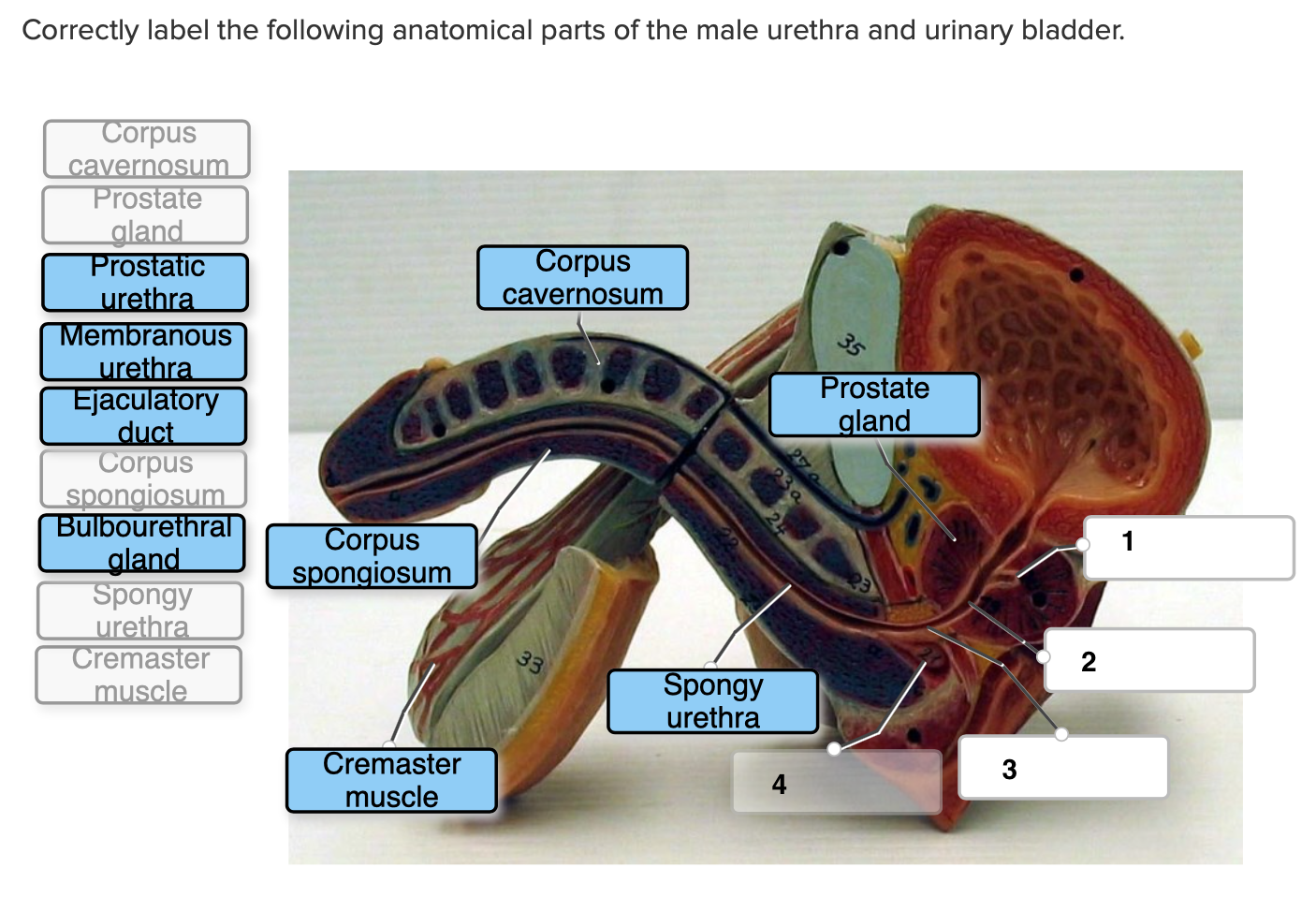
The urethra is the vessel responsible for transporting urine from the bladder to an external opening in the perineum.. It is lined by stratified columnar epithelium, which is protected from the corrosive urine by mucus secreting glands.. In this article, we shall look at the anatomy of the male and female urethra - their anatomical course, neurovascular supply, and any clinical correlations. This page discusses the anatomy and function of the urethra, highlighting differences between males and females. The urethra transports urine from the bladder to the outside of the body for disposal. The urethra is the only urologic organ that shows any significant anatomic difference between males and females; all other urine transport

Medicine LibreTexts Biology Diagrams
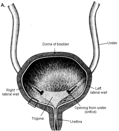
The urinary bladder is a hollow, spherical-shaped organ that holds urine (pee). For most people, it can hold 500-700 mL (about two cups) of pee. When you need to use the restroom, muscles in your bladder contract (tighten) and sphincter muscles in your urethra relax, allowing pee to flow out of your body.

Issues with the urethra are more common in people who have male urethral anatomy. Urinary tract infections, including catheter issues. Urinary tract infections are very common. This test lets your provider look into your urethra and bladder with a cystoscope (a small scope with a camera). X-rays and/or ultrasound. These imaging tests allow
Anatomy of the Urinary System Biology Diagrams
Urinary bladder (sagittal view) The urinary bladder is a pelvic organ that collects and holds urine before urination. It serves as a temporary reservoir for urine produced by the kidneys.When empty, it lies completely within the pelvic cavity, but enlarges upward into the abdominal cavity when full.It is the most anterior pelvic organ, located just behind the pubic bones and pubic symphysis.
